
Here we have the first in a series of posts about lab instrumentation. We in IT know these things as “data makers” but I think its good to understand some of the characteristics behind them and what these machines are actually doing in the way of science. To start, lets talk about Flow Cytometry. A Flow Cytometer is one of the more common instruments used in the lab today. It serves three main functions: a count of the number of cells in a sample, the size / granularity which are generally used together and the identification of cells.
-
The count is the actual number of cells usually counted in the ten’s of thousands
-
The size is counted on a logarithmic scale but the importance is more around the size in relation to other cells in the sample. The cell granularity can be thought of as cell complexity, which is basically the “stuff” inside the cell.
-
The identification of cells is where we learn which cells are exhibiting a particular feature (ie. protein) that the scientist is interested in
All of these are important factors as it will help the scientist determine the next step in their experiment or even if what they’re pursuing is worthwhile.
Now the fun part, LASERS!!!!!!!
The sample is put into the Flow Cytometer and it passes through a “Giant Laser”…er well… not exactly giant, but it does hit each and every cell as they pass through in a single file manner. This is where we get the count. As each cell passes through the laser it causes light to scatter. There are two ways it scatters. We have the forward scatter, which can be correlated to the size of the cell and the side scatter which can be correlated to the granularity of the cell.
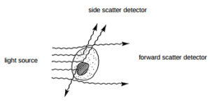
The laser can also detect the presence of different color fluorescents, which can help determine if the cell contains certain features important to overall experiment. As an example, a scientist studying immune response would use fluorescents in flow cytometry to identify a cell specific for a flu virus.
The process is pretty simple, the scientist adds in certain antibodies to the sample that bind to what they are looking for, say a particular protein . These special antibodies “fluoresce” with a specific color (designated by the scientist) to identify the cell as having the antibody, and thus the protein the scientist is looking for. All of this work is done beforehand by the scientist in a manner that they think would work best in the combined cell sample.
For a cool animated 4 min video of how this all works, check this out:
https://www.youtube.com/watch?v=EQXPJ7eeesQ
After the sample is run, the instrument produces a file with a .FCS extension (Flow Cytometry Standard). This file is typically analyzed with a software kit called FlowJo developed by a company with the same name. The scientist would load up FlowJo to view their data, which look something like this:
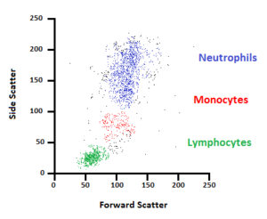
Here you can see the results data on a scatter plot and identify the cell size (forward scatter), the cell granularity (side scatter) and the different cell types.
Now where there is science, there is data, and where there is data, there is an IT person wondering why there is so much of it 🙂
Good news is the output of a flow cytometer is a file size around a few MB, bad news is that in a large lab there can be TONs of these files. The scientists also generate multiple graphs and pictures that are exported into documents, spreadsheets..etc so the downstream effect can be quite large. It’s no where near the amount seen in Next Generation Sequencing or CryoEM (we’ll cover these in another post), but a sizable amount of data in its own right.
For this, a scalable NAS solution such as Isilon is recommended, especially if you think the files are going to be around for a while (hint: they are) you can leverage native cloud tiering to your favorite cloud provider with CloudPools. You also may seen benefits of deduplication as there is a lot of post analysis sharing going on between the PIs (Principle Investigators), their post docs, grad students, technicians..etc.
So there you have it in a nutshell, Flow Cytometry….. and if you remember nothing else, remember this:

Sources:
http://www.bu.edu/flow-cytometry/files/2010/10/BD-Flow-Cytom-Learning-Guide.pdf
https://www.flowjo.com/solutions/flowjo
https://www.labome.com/method/Flow-Cytometry-A-Survey-and-the-Basics.html